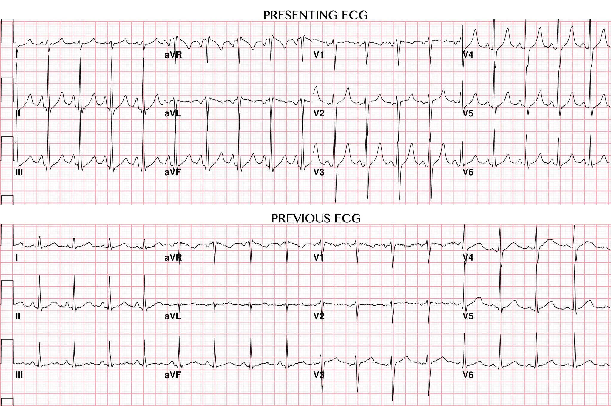

Please share your thoughts on this Patient Case Quiz in the comments section below. Electrocardiographic changes of severe hyperkalemia. The patient's calcium level was 9.2 mg/dl (8.2-10.5 mg/dl). Although the QT interval is slightly short, the T wave is wider. The changes were likely because of a metabolic acidosis due to metformin in concert with hypovolemia, leading to hyperkalemia in the setting of baseline borderline renal function. Tall, symmetric, peaked T waves are visible initially. Slowing of conduction is characterized by an increased PR interval and shortening of the QT interval. ECG changes (see figure ECG patterns in hyperkalemia ECG patterns in hypokalemia ) are frequently visible when serum potassium is > 5.5 mEq/L. Other abnormal blood results include a creatinine level of 6.9 mg/dl (0.5-1.2 mg/dl), a blood urea nitrogen level of 63 mg/dl (7-22 mg/dl), a glomerular filtration rate of 6 ml/min, a bicarbonate (HCO 3) level of 17 mmol/dl (22-30 mmol/dl), and an anion gap of 20 (7-17 mmol/dl). ECG should be done on patients with hyperkalemia.

The patient has a serum potassium level of 7.3 mg/dl (3.5-5.0 mg/dl).
HYPERKALEMIA ECG CHANGES SERIES
A well-defined series of changes at the cellular level leads to characteristic evolutionary changes in the surface electrocardiogram. While the T wave of hyperkalemia is often peaked, it is always narrow, and frequently not peaked. Hyperkalemia is a common cause of electrolyte induced cardiac conduction disturbance. This change is suggestive of hyperkalemia. The T wave is remarkable for the narrow shape due to an abbreviated depolarization time. The patient is in sinus rhythm with normal QRS axis and normal QRS-T complex. T wave changes secondary to hyperkalemia.


 0 kommentar(er)
